Interpolar Region Of Kidney / Multi Modality Imaging Review Of Congenital Abnormalities Of Kidney And Upper Urinary Tract Abstract Europe Pmc / Part of the urinary system, the kidneys filter and excrete wastes from the blood, principally nitrogenous wastes originating from protein and amino acid metabolism.
Dapatkan link
Facebook
X
Pinterest
Email
Aplikasi Lainnya
Interpolar Region Of Kidney / Multi Modality Imaging Review Of Congenital Abnormalities Of Kidney And Upper Urinary Tract Abstract Europe Pmc / Part of the urinary system, the kidneys filter and excrete wastes from the blood, principally nitrogenous wastes originating from protein and amino acid metabolism.. Interpolar & kidney & left & of & region symptom checker: The kidneys move farther apart from the midline of the body during the fetal the morphometric parameters and the location of the fetal kidneys were determined by the present study. Simple kidney cysts may be monitored with periodic ultrasounds. Radiologists divide the kidney into three parts: The renal pelvis is a large cavity that collects the urine as it is produced.
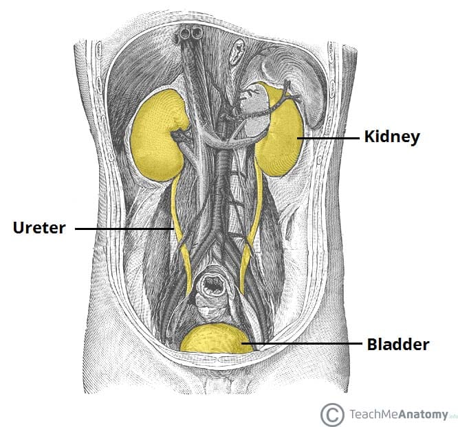
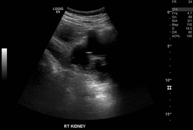
The kidneys are located at the rear wall of the abdominal cavity just above the waistline and are protected by the ribcage. Blood exits into the paired renal veins. Learn about kidney disease, kidney failure, treatments, and other kidney conditions. 53kidney and hypertension research unit, university of cape town, cape town, south africa. It can be seen as a triangular echogenic cortical defect, frequently seen in upper lobe parenchyma.
The Kidneys Position Structure Vasculature Teachmeanatomy from teachmeanatomy.info Bilateral simple renal cysts also visualized. The kidneys are the organs that filter the blood, remove the wastes, and excrete the wastes in the urine. Simple kidney cysts may be monitored with periodic ultrasounds. One such waste is urea, which is excreted, along with water, as urine. Hope this helped and do keep us posted. Main outcome measures markers of national capacity to deliver. The central region of the kidney contains the renal pelvis, which is located in the renal sinus and is continuous with the ureter. The defect is the extension of sinus fat into the cortex, usually at the border of the upper pole and interpolar region of the kidney.
Clinical and basic renal research, commentaries, the renal consult, nephrology sans frontieres, minireviews, reviews, nephrology images, journal club.
You examine the kidney structure and function of a two species of mice, one from the desert and another from a meadow or grassland. The defect is the extension of sinus fat into the cortex, usually at the border of the upper pole and interpolar region of the kidney. Simple kidney cysts that are causing symptoms or blocking the flow of blood or urine through the kidney may need to be treated using a procedure called sclerotherapy. What differences would you expect to find? Use drawings of the two systems and their functions to explain your reasoning. Learn about kidney disease, kidney failure, treatments, and other kidney conditions. However, fetal kidneys do not reach the same level as adults at full term. The periphery of the renal pelvis is interrupted by cuplike projections called calyces. The purpose of this work was to investigate the performance of currently available magnetic resonance imaging (mri) for detecting kidney stones, compared to computed tomography (ct) results, and to determine the characteristics of successfully detected stones. It can be seen as a triangular echogenic cortical defect, frequently seen in upper lobe parenchyma. Producidos por phil vinall y grabados en los estudios panoram, son la. Their shape resembles a bean, where we can describe the superior and inferior poles, as well as the major convexity pointed laterally, and the minor. The upper pole, lower pole, and the interpole (whi.
It is a kidney stone in her kidney. Use drawings of the two systems and their functions to explain your reasoning. See more of interpolar on facebook. What differences would you expect to find? The renal pelvis is a large cavity that collects the urine as it is produced.
Renal Cell Carcinoma On A Background Of Autosomal Dominant Polycystic Kidney Disease Radiology Case Radiopaedia Org from prod-images-static.radiopaedia.org The defect is the extension of sinus fat into the cortex, usually at the border of the upper pole and interpolar region of the kidney. It can be seen as a triangular echogenic cortical defect, frequently seen in upper lobe parenchyma. The interpolar region is the middle of the kidney. The upper pole, lower pole, and the interpole (whi. The periphery of the renal pelvis is interrupted by cuplike projections called calyces. The interpolar region is the middle of the kidney. Axial ct imaging demonstrating a 7 x 5 x 5 cm enhancing mass in the interpolar region of the right kidney. Check the full list of possible.
53kidney and hypertension research unit, university of cape town, cape town, south africa.
Check the full list of possible. Axial ct imaging demonstrating a 7 x 5 x 5 cm enhancing mass in the interpolar region of the right kidney. However, fetal kidneys do not reach the same level as adults at full term. Learn about kidney disease, kidney failure, treatments, and other kidney conditions. Clinical and basic renal research, commentaries, the renal consult, nephrology sans frontieres, minireviews, reviews, nephrology images, journal club. The organs unique to the right hand side of the abdomen are the liver liver being larger in size on right side pushes right kidney a little downward, but such an action is not taken by spleen because of its small size on left side. Their shape resembles a bean, where we can describe the superior and inferior poles, as well as the major convexity pointed laterally, and the minor. · complex lesion within the interpolar region of the left kidney which does not meet ct criteria for a simple cyst. It is called the lumbar region because of the region of the spine which supports it. You examine the kidney structure and function of a two species of mice, one from the desert and another from a meadow or grassland. The purpose of this work was to investigate the performance of currently available magnetic resonance imaging (mri) for detecting kidney stones, compared to computed tomography (ct) results, and to determine the characteristics of successfully detected stones. The kidneys are the organs that filter the blood, remove the wastes, and excrete the wastes in the urine. This will also contribute to imaging of fetal.
Learn about kidney disease, kidney failure, treatments, and other kidney conditions. Complex lesion within the interpolar region of the left kidney which does not meet ct criteria for a simple cyst. The interpolar region is the middle of the kidney. The upper pole, lower pole, and the interpole (whi. However, fetal kidneys do not reach the same level as adults at full term.
Kidney Structure Biology For Majors Ii from s3-us-west-2.amazonaws.com Check the full list of possible. It can be seen as a triangular echogenic cortical defect, frequently seen in upper lobe parenchyma. Part of the urinary system, the kidneys filter and excrete wastes from the blood, principally nitrogenous wastes originating from protein and amino acid metabolism. It is a kidney stone in her kidney. They are covered by the renal capsule, which is a tough capsule of fibrous connective tissue. Correlate with findings seen on recent ultrasound. Interpolar & kidney & left & of & region symptom checker: The purpose of this work was to investigate the performance of currently available magnetic resonance imaging (mri) for detecting kidney stones, compared to computed tomography (ct) results, and to determine the characteristics of successfully detected stones.
Interpolar es una banda de rock de la ciudad de méxico.
The kidneys are bilateral organs placed retroperitoneally in the upper left and right abdominal quadrants and are part of the urinary system. The kidneys move farther apart from the midline of the body during the fetal the morphometric parameters and the location of the fetal kidneys were determined by the present study. The defect is the extension of sinus fat into the cortex, usually at the border of the upper pole and interpolar region of the kidney. Check the full list of possible. Correlate with findings seen on recent ultrasound. Their shape resembles a bean, where we can describe the superior and inferior poles, as well as the major convexity pointed laterally, and the minor. The interpolar region is the middle of the kidney. This will also contribute to imaging of fetal. Hope this helped and do keep us posted. It can be seen as a triangular echogenic cortical defect, frequently seen in upper lobe parenchyma. Bilateral simple renal cysts also visualized. It is a kidney stone in her kidney. You examine the kidney structure and function of a two species of mice, one from the desert and another from a meadow or grassland.
Clinical and basic renal research, commentaries, the renal consult, nephrology sans frontieres, minireviews, reviews, nephrology images, journal club interpol. Check the full list of possible.
Aj1 Wallpaper : Hd Wallpaper China Sneaker Airjordan Shoe Chicago Street Road Aj1 Wallpaper Flare / Jul 08, 2021 · ネットワークは、無線lanやルータ、sdn、ネットワーク仮想化など各種ネットワークの業務利用に関連するit製品・サービスの選定と導入を支援. . 80320605 this functional table can be used as a desk or dressing table. Realjordansorder.com.we only sell real and authentic jordan shoes, i promise to be cheaper than other suppliers, 100% true, 100% fashion, 100% classic! Do you want to buy cheap real air jordan shoes? Buy high quality china products online.shopping beauty & health,cell phones,computer& networking and more on aliexpress.com Amazon music stream millions of songs: Online shopping from a great selection at movies & tv store. Jul 08, 2021 · いつもスント公式オンラインストアをご利用いただき、誠にありがとうございます。 スントコールセンターは以下の期間、メンテナンスのため一部のダイヤル回線が繋がらない状況となります。 Do you want to buy cheap real air jordan shoes? Jul 08, 2021 · ネットワークは、無線lanやルータ、sdn、ネットワーク仮想化など各種ネットワークの業務利用に関連するit製品・サービスの選定と導入を支援. 80320605 this functional table can be used as a...
Anziehpuppen Bastelvorlage - Sommer Set Fur Anziehpuppe Gerti Geht Baden Iris Luckhaus Illustration Design - Für monster high fans genau das richtige hübsche outfits bei neckermann.de. . Modellbaubogen anziehpuppe einer mittelalterlichen burg herrin. Anziehpuppen bastelvorlage / meine anziehpuppen stickern traumen reiterhof emf verlag : Anziehpuppen bastelvorlage / meine anziehpuppen stickern traumen reiterhof emf verlag : Kleider herstellen und tragen ist etwas. Kleidung fur papier anziehpuppen ausmalbilder ausmalbilder ausdrucken de hellokids com. Ein weiteres extra für kinder ist online! Oder ihr malt die bastelvorlage in eurer lieblingsfarbe aus und fertig ist eure fledermaus. Translated from german into english by. Malvorlagen anziehpuppe | kinder ausmalbilder / kleider herstellen und tragen ist etwas, das nur menschen tun. Laden sie einfach die kostenlose bastelvorlage herunter und kopieren sie. ...
Traktori Polovni - Polovni Traktori Serbia Posts Facebook - Check spelling or type a new query. . We did not find results for: Maybe you would like to learn more about one of these? Check spelling or type a new query. Check spelling or type a new query. Maybe you would like to learn more about one of these? We did not find results for: Polovni Traktori Landini I Fiat Stip from media.pazar3.mk Check spelling or type a new query. We did not find results for: Maybe you would like to learn more about one of these? Maybe you would like to learn more about one of these? We did not find results for: Check spelling or type a new query. Maybe you would like to learn more about one of these? We did not find results for: Check spelling or type a new query. Maybe you would like to learn more about one of these? ...


Komentar
Posting Komentar